Nwredhead
Gallery · April 23, 2025
Ct Brain Anatomy Tutorial
Ct Brain Anatomy Tutorial BASIC APPROACH TO EVALUATING A HEAD CT
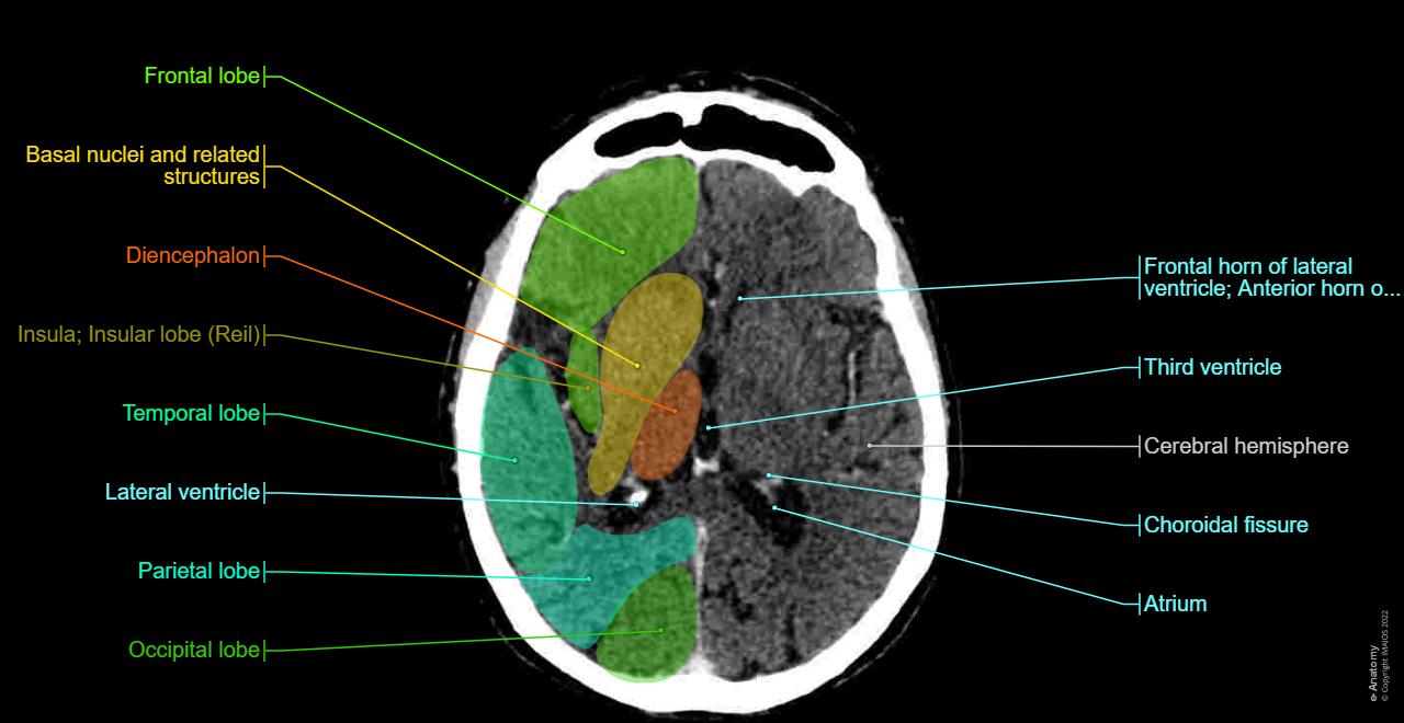
Anatomy of the brain and face: labeled CT - e-Anatomy
image size: 1280x660

Anatomy of the brain and face: labeled CT - e-Anatomy
image size: 1280x660

Anatomy of the brain and face: labeled CT - e-Anatomy
image size: 1280x660

Anatomy of the brain and face: labeled CT - e-Anatomy
image size: 1280x660

Anatomy of the brain and face: labeled CT - e-Anatomy
image size: 1280x660

Anatomy of the brain and face: labeled CT - e-Anatomy
image size: 1280x660

CT scan of normal brain anatomy
image size: 1440x1615

CT Angio Atlas | neuroangio.org
image size: 1094x735

Understanding Brain CT Scans in Neurosurgery
image size: 1233x1764

CT Angio Atlas | neuroangio.org
image size: 1095x739

Normal CT Anatomy of the Brain https://www.youtube.com/watch?v\u003dQ9RoYxtuc7Q\u0026t\u003d262s In this video, we will be going through CT axial slices of a normal brain. Thank you to Radiopedia for the wonderful CT scan images. #
image size: 1536x1024

CT scan of normal brain anatomy
image size: 1772x1617

Head CT Anatomy and Interpretation | PDF | Wellness | Science \u0026 Mathematics
image size: 768x1024

Normal CT Anatomy of the Brain https://www.youtube.com/watch?v\u003dQ9RoYxtuc7Q\u0026t\u003d262s In this video, we will be going through CT axial slices of a normal brain. Thank you to Radiopedia for the wonderful CT scan images. #
image size: 960x950

CT scan of normal brain anatomy
image size: 2048x2048

How to Read a CT Scan of the Head - MEDZCOOL
image size: 1280x720

How to perform a brain CT perfusion coregistration with anatomic atlas? - Software Support - Neurostars
image size: 875x973

Understanding CT Brain Anatomy for Medical Students
image size: 1080x1920

BASICS of CT Head | PPTX
image size: 2048x1536

Med Students | Navigating Radiology
image size: 1280x720

CT Head Anatomy: Understanding the Basal Ganglia and Cisterns
image size: 884x1920

CT Anatomy \u0026 Procedures Guide | PDF | Human Brain | Corpus Callosum
image size: 768x1024

BASICS of CT Head | PPTX
image size: 2048x1536

brainstorm_sEEG
image size: 2236x2092

Fully Automated Segmentation of Head CT Neuroanatomy Using Deep Learning | Radiology: Artificial Intelligence
image size: 1700x2181

Midbrain, Pons, and Medulla: Anatomy and SyndromesRadioGraphics
image size: 3200x2197

How to perform a brain CT perfusion coregistration with anatomic atlas? - Software Support - Neurostars
image size: 2360x791

MRIcroGL Tutorial #4: Viewing Results — Andy's Brain Book 1.0 documentation
image size: 2578x1024

Mediphany | How to read an MRI or CT scan
image size: 1564x953

emergency radiology — Blog — NUEM Blog
image size: 1184x727

Spot Diagnosis on CT Brain | Medical Student's Guide
image size: 750x1334

Normal CT Anatomy of the Brain https://www.youtube.com/watch?v\u003dQ9RoYxtuc7Q\u0026t\u003d262s In this video, we will be going through CT axial slices of a normal brain. Thank you to Radiopedia for the wonderful CT scan images. #
image size: 1536x2048

CT Brain interpretation | PPTX
image size: 2048x1536

CT Brain Scan Explained: Identify Key Abnormalities
image size: 1014x1473

Ischemic stroke | Radiology Reference Article | Radiopaedia.org
image size: 1280x720

Head CT — Blog — NUEM Blog
image size: 2500x1608

How To 3D Print Your Brain In A Few Simple Steps - TUTORIAL 2022
image size: 1280x720

Head CT vs Brain MRI: Understanding the Key Differences
image size: 1080x1920

Generative adversarial network–based reconstruction of healthy anatomy for anomaly detection in brain CT scans
image size: 1620x963

CT Scan of the Brain Without Contrast: A Protocol Guide for CT Techs
image size: 1280x720

CT Anatomy of Ear | enteducationswansea
image size: 3685x1648

brainstorm_sEEG
image size: 3380x1811

Voxel-Based Morphometry with CAT12 — Andy's Brain Book 1.0 documentation
image size: 1772x2568

Foundational features of the brainstem
image size: 1280x720

Basic Principles of CT Scans - Interpretation - TeachMeAnatomy
image size: 1600x1100

CT Anatomy of Ear | enteducationswansea
image size: 3677x1648

Visualizing MRI \u0026 CT Scans in Mixed Reality / VR / AR, Part 2: 3D Volume Rendering – andreasjakl.com
image size: 1920x1040

Brain MRI scan protocols, positioning and planning
image size: 1280x720

CT Brain Imaging Basics and Techniques | PDF | Ischemia | Cerebellum
image size: 768x1024

Head CT — Blog — NUEM Blog
image size: 1600x4228

Vascular | Learn Neuroradiology
image size: 1280x720

Visualizing MRI \u0026 CT Scans in Mixed Reality / VR / AR, Part 2: 3D Volume Rendering – andreasjakl.com
image size: 2000x1125

AAPM/RSNA Physics Tutorial for Residents: Topics in CTRadioGraphics
image size: 1800x1401

Cerebral circulation 1: anatomy - BJA Education
image size: 1476x1194

Head CT — Blog — NUEM Blog
image size: 1600x4005

Anatomy of the Heart and Great Vessels on CT
image size: 1280x720

Med Students | Navigating Radiology
image size: 1280x720

Three‐Dimensional Cinematic Rendering to Optimize Visualization of Cerebrovascular Anatomy and Disease in CT Angiography - Caton - 2020 - Journal of Neuroimaging - Wiley Online Library
image size: 900x1011

Unit 1: Neurons \u0026 Brain Anatomy | The Franklin Institute
image size: 1620x1135

Imaging of Stroke: Part 1, Perfusion CT???Overview of Imaging Technique, Interpretation Pearls, and Common Pitfalls | AJR
image size: 1710x1710

I put my brain into Blender! (More info in comments) : r/blender
image size: 1920x1080

Head CT — Blog — NUEM Blog
image size: 1600x6585

brainstorm_sEEG
image size: 3380x1811

Issue with Head Modeling - Discussions - Brainstorm
image size: 912x1126

An integrated teaching method of gross anatomy and computed tomography radiology - Murakami - 2014 - Anatomical Sciences Education - Wiley Online Library
image size: 1204x756

Vascular | Learn Neuroradiology
image size: 1280x720

Generative adversarial network–based reconstruction of healthy anatomy for anomaly detection in brain CT scans
image size: 1621x541

CT Anatomy of Ear | enteducationswansea
image size: 3674x1648

Brain Clipart, Brain Anatomy Graphic, Brain Clip Art, Anatomical Brain, Brain Anatomy Art, Medical Illustration, Neurology, Neurologist Art - Etsy
image size: 2000x1600

Cervical spine CT (computed tomography) radiology search pattern
image size: 1280x720

Unit 1: Neurons \u0026 Brain Anatomy | The Franklin Institute
image size: 1620x1135

CAT12 Manual
image size: 1999x1125

Imaging of Stroke: Part 1, Perfusion CT???Overview of Imaging Technique, Interpretation Pearls, and Common Pitfalls | AJR
image size: 1800x1800

Unit 1: Neurons \u0026 Brain Anatomy | The Franklin Institute
image size: 1280x720

Neuroradiology threads by Lea Alhilali - Functional brain anatomy | Radiopaedia.org
image size: 1200x684

Beginner question: liver volumetry - Support - 3D Slicer Community
image size: 1920x1005

3d Ct Scan Brain Showing Mass Stock Illustration 2646772409 | Shutterstock
image size: 1429x1600

MRIcroGL Tutorial #4: Viewing Results — Andy's Brain Book 1.0 documentation
image size: 2166x1114

CT Brain Posterior Fossa Anatomy | PDF | Science \u0026 Mathematics | Computers
image size: 768x1024

MR Cranial Bone Imaging: Evaluation of Both Motion-Corrected and Automated Deep Learning Pseudo-CT Estimated MR Images | American Journal of Neuroradiology
image size: 1280x1166

POCUS/Imaging – critical care notes
image size: 1280x720

How-To: Creating a 3D Reconstruction of Your Patient's CT Scan
image size: 1280x720

Neurology Clinical Rotation: Week in the Life of a Year 3 Medical Student - The Lowkey Medic
image size: 1200x801

One-Minute CT Preprocessing with Miptools | by Enoch Kan | TDS Archive | Medium
image size: 1280x960

Bringing Radiology Education to a New Reality: A Pilot Study of Using Virtual Reality as a Remote Educational Tool - Yuhao Wu, Prosanta Mondal, Matthew Stewart, Richard Ngo, Brent Burbridge, 2023
image size: 2980x1966

Imaging Tutorial: Differential Diagnosis of Bright Lesions on Diffusion-weighted MR ImagesRadioGraphics
image size: 1800x1800

3d Ct Angiography Circle Willis Human Stock Illustration 2672043205 | Shutterstock
image size: 1500x1519

00P-public-course-page
image size: 1920x1080
Cross sectional anatomy | Kenhub
image size: 1400x896

Three‐Dimensional Cinematic Rendering to Optimize Visualization of Cerebrovascular Anatomy and Disease in CT Angiography - Caton - 2020 - Journal of Neuroimaging - Wiley Online Library
image size: 900x885

MR Cranial Bone Imaging: Evaluation of Both Motion-Corrected and Automated Deep Learning Pseudo-CT Estimated MR Images | American Journal of Neuroradiology
image size: 1145x1280

Arterial Supply to the Brain - Carotid - Vertebral - TeachMeAnatomy
image size: 1688x626

Clover Learning launches CT anatomy courses | Ari Blum posted on the topic | LinkedIn
image size: 1273x720

Frontiers | Development and validation of a prognostic computed tomography scoring model for functional outcomes in patients with large hemispheric infarction following decompressive craniectomy
image size: 1898x920

Brain CT Anatomy and Basic Interpretation Part I | PPT
image size: 2048x1536

Terminology and Anatomy of the Nervous System – Glass Box Medicine
image size: 1200x675

Automatic whole-body CT segmentation in 2 minutes using 3D Slicer and TotalSegmentator
image size: 1280x720

A deep learning method for automatic segmentation of the bony orbit in MRI and CT images | Scientific Reports
image size: 2029x896

BASIC APPROACH TO EVALUATING A HEAD CT
image size: 1920x1050
Discover more galleries
- August 2025 Tim Robbins Filme /2025-08/tim-robbins-filme
- February 17, 2025 Shoulder Jewelry Accessories /2025-02-17/shoulder-jewelry-accessories
- October 26, 2025 Wallpaper Love Quotes For Him /2025-10-26/wallpaper-love-quotes-for-him
- June 2025 Ballroom And Latin Quotes /2025-06/ballroom-and-latin-quotes
- April 12, 2025 Energia Nuclear Ejemplos /2025-04-12/energia-nuclear-ejemplos
- January 3, 2025 Hickory Woods Golf Course Map /2025-01-03/hickory-woods-golf-course-map
- May 2025 Brunette Men /2025-05/brunette-men
- July 10, 2025 American Girl Catalog July 2017 /2025-07-10/american-girl-catalog-july-2017